Ovarian cancer rarely produces symptoms until advanced stages. This pathology image shows why: by the time it is finally removed, the tumor already contains multiple cell populations.
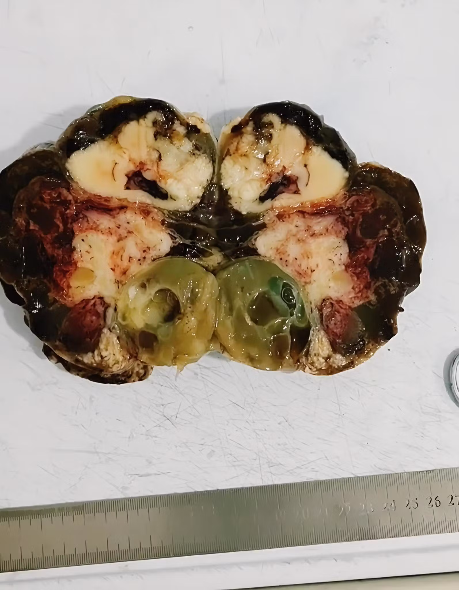
On cross-section, at least three morphologically distinct zones can be identified. The peripheral region exhibits dark hemorrhagic tissue with frank necrosis, a sign that the tumor vascularization collapsed before the tissue could be operated on. In the lower center, a cystic structure with olive-green mucinous content, characteristic of mucinous cystadenocarcinomas, the histological subtype that can reach 30 centimeters in diameter without causing pain.
The whitish central zone corresponds to undifferentiated solid tissue. The millimeter ruler in the image indicates that the specimen exceeds 25 centimeters along its greatest axis. Ovarian cancer has a five-year survival rate of 49% in late stage, precisely because this architectural complexity causes it to respond heterogeneously to chemotherapy.